
Using the same metal for both the grid and the foil improves the quality of your data because it reduces beam-induced motion caused by changes in the geometry and tension of the foil due to the elimination of the differential thermal contraction between grid and metal during plunge vitrification and sample irradiation. As discussed in Russo and Passmore, Science 346: 1377-1380 (2014) and Russo and Passmore. J. Struct. Biol. 193: 33-44 (2016).
-
Our products
-
View all products
-
HexAuFoil QUANTIFOIL® Holey Carbon Supports for Cryo-EM UltrAuFoil® Holey Gold Sample Supports Additional Ultrathin Continuous Carbon Layer Conventional & Continuous Carbon Films SiO2 Films Custom Cryo-EM Sample Supports and Special Treatments QUANTIFOIL® Active How our supports are packed for shipment
-
-
View all products
- Our company
- News and social updates
- Support
UltrAuFoil®
Better 3D reconstructions from less data with ultra-stable gold supports for cryo-electron microscopy that reduce the movement of frozen specimens during imaging.
Designed at MRC's Laboratory of Molecular Biology by Dr Christopher J Russo and Dr Lori A Passmore and manufactured exclusively under license by Quantifoil Micro Tools GmbH1, UltrAuFoil® Holey Gold sample supports make structure determination for challenging and small molecules easier.
During imaging at cryogenic temperatures, traditional carbon supports move, particularly at the beginning of irradiation. This movement blurs images and reduces data quality. UltrAuFoil® Holey Gold supports are more conductive, and there is no differential contraction between the grid and the foil on plunge freezing. Therefore there is less crinkling of the foil during sample preparation, resulting in many improvements in data quality.
UltrAuFoil® Gold supports for better reconstructions with less data
- Retains your highest resolution data, before the sample accumulates radiation damage, by facilitating the use of the first frames in your movies due to reduced beam-induced motion.1,2
- Increases resolution, by up to 0.5 Å when replacing traditional holey carbon supports with UltrAuFoil®.3
- Reduces image distortion from the accumulation of static and semi-mobile charge as gold is highly conductive at liquid nitrogen temperatures.1
- Protects samples from damage due to accumulating positive charge by neutralizing it with secondary electrons generated by irradiating adjacent gold.1
- Allows biomolecules to retain native structure and by not putting them under mechanical strain from foil crinkling.
- Improves particle distribution as the gold foil is even flatter than carbon, and capable of forming the <200 Å ice layers required for ultra-high resolution data collection on smaller molecules.4
- Simplifies grid surveying due to the high contrast of the gold foil.1
Citations
Since their launch in 2015, UltrAuFoil® holey gold sample supports have become recognized as the optimum choice to maximize reconstruction quality and resolution. They are now referred to in approximately 500 peer-reviewed publications.
Reviews and best practice
A number of recent publications describing best practice for sample preparation for cryoEM data collection refer to the use of UltrAuFoil® holey gold films, including:
Chua et al. Better, Faster, Cheaper: Recent Advances in Cryo–Electron Microscopy. Ann. Rev. Biochem. 91: 1-32 (2022)
Danev et al. Routine sub-2.5 Å cryo-EM structure determination of GPCRs. Nat. Commun. 12: 4333 (2021)
Passmore and Russo. Specimen Preparation for High-Resolution Cryo-EM. Methods Enzymol. 579: 51-86 (2016)
Examples of UltrAuFoil® grids in recent SPA structure solutions
Greenhough et al. Structure and function of the RAD51B-RAD51C-RAD51D-XRCC2 tumour suppressor. Nature 619:650-657 (2023)
Meier et al. Structural and functional characterization of the Sin Nombre virus L protein. PLoS Pathog 19: e1011533 (2023)
Grba et al. Investigation of hydrated channels and proton pathways in a high-resolution cryo-EM structure of mammalian complex I. Sci. Adv. 9: eadi1359 (2023)
Other applications
Holey gold films have reduced fluorescence compared to standard Carbon foils, and are biocompatible. Therefore, they are can provide benefits for tomography and correlative studies. Recent examples include:
Dumoux et al. Cryo-plasma FIB/SEM volume imaging of biological specimens. Elife 12: e83623 (2023)
Dahmane. Membrane-assisted assembly and selective secretory autophagy of enteroviruses. Nat. Commun. 13: 5986 (2023)
You can discover more about how UltrAuFoil® holey gold foils have been used by researchers in our list of peer-reviewed publications.

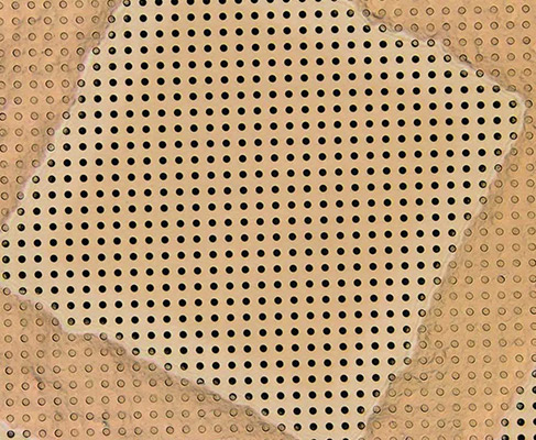
Characteristics of UltrAuFoil®
Thickness of gold foil
About 500 Å
Foil geometries and grid specifications
R 0.6/1 and R 1.2/1.3 Gold foil on Gold 300 mesh grid
R 2/2 Gold foil on Gold 200 mesh grid
Other combinations on request
Frequently asked questions
UltrAuFoil® are an exciting development in specimen support technology. As many customers would like to know more about them, we've gathered together some of the most frequently asked questions below, but if there's anything you're still not sure of please ask us or your local distributor - we're happy to help.
Gold has several properties that make it ideal as a sample support in electron microscopy, including being conductive, non-oxidizing, radiation-hard. In addition, it is does not interfere with the specimens you are studying as it is chemically inert and biocompatible, as described in Russo and Passmore, 2014 and 2016.
A foil thickness of 400-500 Å provides optimum data quality by balancing the need for the thinnest possible layer of ice with sufficient thickness of the gold foil to minimize beam-induced motion.
A thicker foil will not further reduce beam-induced motion, but risks degrading data quality due to thicker ice resulting in poor particle dispersion and increased background scatter. In contrast, a thinner foil might offer better particle dispersion due to the formation of a thinner layer of ice, but at <400 Å, the foil is thinner than the individual gold nano-particles it is made from and this increases beam-induced motion due to poorer conductance as the surface becomes uneven. See Russo and Passmore, 2014 and 2016 for further details.
As with our other products, UltrAuFoils® should be stored in a grid storage box in a cool, dark, low-humidity environment. While there is no date of expiry for the UltrAuFoils® we generally recommend using them within two years.
UltrAuFoil® sample supports are ready to use straight from the box. However, as with all transmission electron microscopy grids, users may achieve better sample dispersion and improved wetting if the foils are made more hydrophilic. This can be achieved using standard glow discharge and plasma systems, as described here. UltrAuFoil® grids particularly benefit from these treatments, as, lacking the more volatile carbon foil, they can be safely exposed to extended glow discharge or plasma treatment without risk of any surface degradation.
Yes. We are happy to supply UltrAuFoil® with an ultrathin carbon layer at a thickness of your choice (most commonly 2 nm, but 3, 5 and even 10 nm additional carbon layers are available on request). Alternatively, you may add an additional layer of amorphous carbon yourself: standard float transfer methods, as described in Passmore and Russo, 2016, are recommended to transfer thin films of carbon onto UltrAuFoils®.
Data collection can be carried out as you would for holey carbon foils with similar geometries. Thus, we recommend that the electron beam geometry is circularly symmetric and centered on the hole. The micrograph is taken at the center of the hole. We would recommend including a small section of the support foil in each image, as this aids with focussing (see "How do I focus using UltrAuFoils®", below)
Since there is no amorphous material in the gold support structure, Thon rings cannot be used to focus. Russo and Passmore, 2016, present a number of different options for focussing when using UltrAuFoils®, but the two simplest are:
- Oscillate the beam tilt around 0°, and the plane for which the image shift is minimized is the in-focus setting. This method is most useful for automated data collection.
- The micro-crystals of gold in the foil diffract elections, with the diffraction spots occurring in a ring around the crystal image at a distance related the lattice spacing of the gold crystals, as predicted by the Bragg equation, and the defocus. As the smallest lattice spacing of the Gold crystals gives rise to diffraction at a resolution of 2.35 Å, care should be taken to ensure that any objective aperture does not block electrons at the diffracted frequencies. This method is primarily recommended for manual data collection modes.
We recommend using a calibration specimen to correct the stigmation and beam tilt prior to collecting data on UltrAuFoils®.
Yes, automated data collection has been successfully tested on UltrAuFoils® using beam tilt to focus.
No, they should be handled in the same way as traditional carbon foils, and are similarly robust. As with traditional carbon foils, care should be taken when handling the foils with tweezers and during plunge freezing, as if the foil is damaged in these processes, the stability of the support may be severely degraded. We recommend collecting data only from squares where the foil is uniform and intact.
In addition, the gold foil is not volatile when glow discharged or treated with plasma, so the grids may be subjected to far more extensive plasma treatments than standard carbon foils, without any risk of degrading the surface.
Dr Christopher J Russo and Dr Lori A Passmore of the MRC Laboratory of Molecular Biology, Cambridge, UK invented these sample supports and they are produced under license by Quantifoil Micro Tools, GmbH.
Chris Russo and Lori Passmore have written two very informative papers about the characteristics of these grids and how they compare to more traditional holey carbon foils.